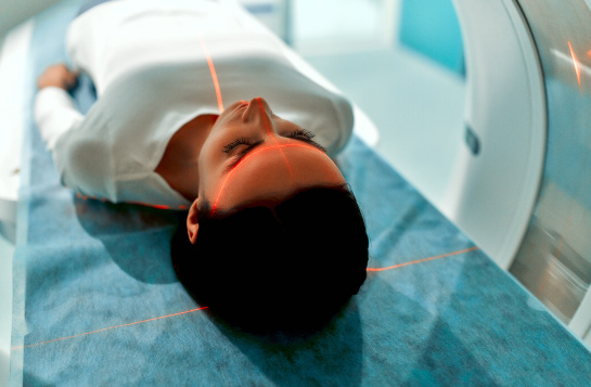
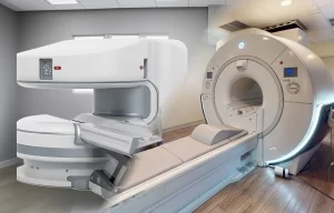
Positron Emission Tomography (PET) scans offer several benefits, making them a valuable tool in medical diagnostics. This non-invasive and painless imaging method allows physicians to detect diseases at early stages and assess areas of activity inside the body.
Non-Invasive:
A non-invasive PET scan utilizes a small quantity of radioactive tracer to observe the blood flow and functionality of the heart muscle tissue. To produce images of the heart at rest and when the doctor gives medication to the patient, a tracer is injected into a vein in the arm and then detected by unique cameras. This detection aids in diagnosing certain heart conditions and specific types of cancer.
The PET scan is a non-invasive and generally painless procedure. A small dose of radioactive material, typically glucose or similar compounds, is administered through a vein, and a specialized positron scanner captures detailed information about the body’s metabolism of the tracer. This imaging method has been in clinical use since the early 1990s.
Painless:
A Painless PET scan involves injecting a radioactive FDG (fluorodeoxyglucose) into the body through a small plastic tube. The patient remains on the scan table for 30 to 60 minutes, and the FDG travels through the blood to the targeted area, where the tissues absorb it. The scan then takes place, providing detailed views of the body and its organs. PET scans help determine organ function by measuring tissue metabolism and effectively detecting brain tumors and heart disease.
The procedure of PET scans is relatively painless, involving the injection of a tracer liquid into a vein, sometimes accompanied by swallowing or inhaling the tracer. As the liquid travels through the body, it emits positrons, which a camera records and transforms into images on a computer screen. PET scans can assess brain trauma, measure perfusion in the heart muscle, and detect the spread of heart disease.
Early Disease Detection:
A PET scan utilizing a special camera and tracer can detect cellular changes in the body, enabling the diagnosis of heart, lung, and brain conditions. Compared to other imaging tests, PET scans excel at detecting diseases in their earliest stages.
Positron emission tomography relies on the physical properties of isotopes that release positrons as they decay. The radioactive substances used in PET scans travel through the bloodstream and bind with brain cells, releasing positrons detected by a pair of specialized detectors. This technique effectively pinpoints diseases at their early stages, aiding in timely diagnosis and treatment planning.
Detects Radioactive Glucose:
PET scans can effectively detect cancer by measuring the levels of radioactive glucose in the body. The procedure involves using a small amount of radioactive glucose, a tracer, to create computerized images
of the body’s chemical structures. PET scans can identify tumors and abnormalities and are frequently combined with CT scans to improve precision.
Preparing for a PET scan typically involves avoiding food, drink, and strenuous exercise for several hours before the procedure to maintain stable sugar metabolism. Patients should wear loose-fitting clothing and remove metal jewelry before the scan. The procedure takes 30 to 60 minutes, during which the patient receives a contrast infusion. After the scan, patients can typically resume their daily activities, and results are usually available within a week.


