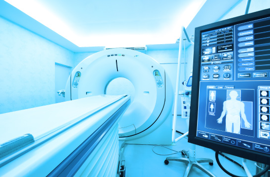
Medical imaging encompasses a diverse array of techniques designed to peer into the depths of the body’s interior, including cutting-edge modalities like X-rays; there are various types of medical imaging techniques available such as Computed Tomography (CT), Magnetic Resonance Imaging (MRI), sonography, and others. Each of these cutting-edge methodologies boasts distinctive advantages, empowering medical practitioners to diagnose intricate conditions and acquire exquisitely detailed images that traditional X-rays might fail to reveal.
X-rays
The realm of X-rays is astonishingly multifaceted and forms a cornerstone of medical imaging practices. By skillfully channeling radiation through the body, X-rays unveil images that facilitate healthcare professionals in gaining unprecedented insights into the inner structures. Introducing a contrast medium occasionally comes into play, meticulously highlighting areas of particular interest. Fortuitously, X-rays have garnered a reputation for safety, meticulously regulated by stringent laws and guidelines to uphold patient welfare. While exposure to ionizing radiation is generally meager, engaging in dialogues with healthcare providers about potential risks remains crucial, primarily if multiple X-ray sessions are necessitated throughout a lifetime. Pregnant women, in particular, are prudently advised against holding their babies during an X-ray session, prioritizing utmost caution. With the advent of pioneering techniques like Single-Frame X-ray Tomosynthesis (SRS), promising augmented resolutions and potential therapeutic efficacy in addressing lung tumors and cardiovascular maladies have become a distinct possibility.
MRI
MRI scans, being a familiar territory, exude an air of safety and reliability, yet patients are advised to undertake certain precautions. At the crux of MRI lies a formidable electromagnet, orchestrating the alignment of hydrogen atoms within the body to unleash signals that, in turn, contribute to the creation of astounding images. Pregnant women are judiciously counseled to warn MRI technologists that applying certain contrast materials could pose hazards to unborn offspring. MRI emerges as a veritable diagnostic powerhouse, unraveling the mysteries of various conditions, such as herniated spinal discs, multiple sclerosis, and liver cirrhosis, while furnishing high-fidelity images and precise data. To ensure a seamless process, patients are required to divest themselves of any metallic objects and observe utmost stillness during the procedure, which typically extends between 15 minutes and an hour.
Sonography
Sonography, basking in the safe embrace of ultrasound waves, emerges as a non-invasive marvel for visualizing internal structures without the perils of radiation or invasive interventions. As the skillful sonographer glides the transducer across the patient’s skin, a captivating tableau of images manifests on the computer screen, unveiling the intricacies of the body’s innermost secrets. Sonography boasts a vast spectrum of applications, encompassing vascular, obstetric, gynecologic, abdominal, musculoskeletal, and cardiovascular imaging. It is a vital tool for diagnosing pelvic masses, infertility challenges, and inflammatory ailments. It offers a realm of promise through its capacity to reveal soft tissue, and bone surfaces, identify muscle strains, ligament sprains, and fractures, and even facilitate needle-guided injections. Adroitly trained sonographers, wielding sophisticated equipment with finesse, navigate their interactions with patients, brimming with expertise in anatomy, communication, and self-assurance throughout the entire procedure. Notably, the real-time capabilities of sonography shatter the shackles of radiation exposure associated with X-rays, heralding a safer alternative replete with a bounty of benefits.


