
For years, a dark shadow has hung over contact sports: Chronic Traumatic Encephalopathy, or CTE. It’s a brain disease caused by repeated head hits. The biggest problem with CTE? Doctors could only diagnose it for sure after a person died, by looking closely at their brain tissue under a microscope.
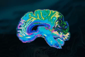
But that is finally starting to change. Thanks to advanced MRI scanning and major university research, scientists are finding ways to spot structural warning signs of CTE in living patients.
Here is a look at the major MRI breakthroughs giving athletes and their families new hope.
Discovering Hidden Brain Grooves at NYU Langone
In late 2025, researchers at NYU Langone Health made a massive discovery using structural MRI. They wanted to know if the brains of former football players looked different from the brains of people who never played contact sports. To find out, the study compared MRI scans from 169 former college and professional football players against 54 healthy men of similar age and education who had never played collision sports.
By looking closely at the images, they found a clear difference. The football players had noticeably shallower grooves on the top left side of their brains. This specific area is called the left superior frontal sulcus, and previous studies show it is heavily affected by Chronic Traumatic Encephalopathy (CTE). The researchers also found that players with longer football careers had wider grooves in another part of the brain. This means that the more years a person plays, the more their brain structure might physically change. Finding these structural changes in living people gives doctors concrete signs to look for on a scan. In the future, this could help doctors identify who is at high risk for CTE while they are still alive.
Using Past Scans to Understand CTE
Boston University (BU) is one of the leading centers for CTE research in the world. They used a clever “look-back” method to figure out what CTE looks like on an MRI. Usually, scientists have to track patients for decades to study a progressive brain disease. To speed up the process, researchers worked backward. They gathered old MRI scans from 55 men who had later died and were confirmed to have CTE. These MRIs were taken an average of four years before the patients passed away.
The researchers compared these scans to MRIs from 31 healthy men. By matching the historical MRIs with the physical brain exams done after death, they discovered a distinct pattern. Brains with confirmed CTE showed measurable shrinkage in the frontal and temporal lobes, which are the main areas damaged by the disease. The researchers also found that people with CTE were almost seven times more likely to have a specific brain abnormality called a cavum septum pellucidum (CSP), which is a tear or space near the center of the brain. This research brings doctors much closer to diagnosing CTE in living patients using standard MRI machines.
Inside the DIAGNOSE CTE Project
The multi-site DIAGNOSE CTE Research Project, which includes major medical hubs like the Cleveland Clinic, is another driving force. This extensive study relies heavily on structural MRI scans and neurological assessments to evaluate former athletes (such as football players and fighters) who show clinical symptoms of Traumatic Encephalopathy Syndrome (TES).
By mapping brain volumes through MRI during these comprehensive multi-day evaluations, researchers can track tissue loss and structural changes over time. This helps map out how repetitive head impacts alter key areas of the brain while patients are still alive.
What This Means for the Future
Although MRI alone cannot provide a standalone, definitive label for CTE. However, these university breakthroughs prove that advanced structural imaging is becoming an indispensable tool.
As researchers combine MRI brain volume tracking with supplemental blood tests to flag injury proteins, the goal of diagnosing and managing CTE in living patients is closer than ever before.
Tesla MRI utilizes these technologies together for patients at our centers, providing comprehensive brain imaging that improves diagnosis accuracy and enables personalized treatment monitoring in a single efficient visit. Interested in visiting our centers? Contact us at (833) TESLA-MRI.